Patient came in complaining of foreign body sensation in the left eye. Eyelid eversion revealed a mucous excretion from a cyst. Removing the mucous left an interesting peek into the cavern of the cyst. Pressure drained the cyst entirely.
Search Eye Pictures
Thursday, October 8, 2015
Wednesday, September 30, 2015
LASH STUCK IN PUNCTAL OPENING
This patient complained that for weeks his eye felt like it had something in it. Upon examination a loose eyelash had lodged itself in the lower lid punctal opening.
Tuesday, August 25, 2015
HZV KERATITIS
This patient was complaining of extreme pain in and around the eye. HZV usually doesn't affect the cornea but this patient had primary corneal HZV. We were able to catch it really early and get her on the anti-virals very quickly to prevent worsening or lengthening of the manifestations of the disease. Usually HSV does not cause much pain. There were also some iritis associated with this so steroids were added as soon as possible along with the antivirals with success, but this has to be monitored extremely closely.
DIFFUSE CORNEAL SUBEPITHELIAL INFILTRATE
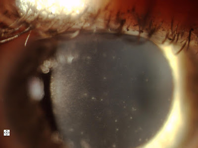
This is a patient complaining about red watery eyes, one eye worse than the other. This photo shows diffuse subepithelial infiltrate associated with punctate staining at each point. The other eye was only about a third as bad. This is adenoviral keratoconjunctivitis.
Wednesday, August 5, 2015
Tuesday, August 4, 2015
CENTRAL SEROUS RETINOPATHY
Central Serous Retinopathy (CSR) occurs when there's serous leakage from the blood-vessel-filled choroid layer of the eye in the space under the retina. This patient's vision was brought from 20/20 in April to 20/40 in August. CSR recovers usually resolves and the vision recovers within a few months, however it is not unusual to have some remaining small deficit in vision. It usually occurs in one eye only but may occur in both, and it may recur.
Tuesday, April 14, 2015
RETINOSCHISIS
You can see the smooth base of the retinoschisis dome and if you enlarge it you can see the retinal blood vessels over the top of the dome.
KERATOCONUS
The slit beam accentuates the irregular curvature of the cornea. The apex of the cone is inferior to the visual axis and you can also see co-existent thinning of the cornea over the apex of the cone.
Subscribe to:
Posts (Atom)


