Search Eye Pictures

Wednesday, December 31, 2008

MYELINATED NERVE FIBERS


Myelin is the insulation around the nerves in our bodies. This myelin helps the electric signal to travel down the nerve in greater efficiency. As the nerve fibers enter the eye, however, they lose their myelin. Sometimes the myelin continues into the retina as an interesting anomoly. Usually the myelin is near the optic nerve. But in this case, there is myelin around the nerve fibers in the peripheral retina, as you can see in the photo. This myelin has no consequence to this patients vision and there is no danger in this small extent.

MYELINATED NERVE FIBERS


This is the optic nerve of a young man. The optic nerve is normally orangish and round. But you can see there's some white material just below this optic nerve (it's actually above since the image is reversed in the photo). You can see the retina blood vessels overlying the white material. This material is "myelination". Myelin is the insulation of the nerves in our bodies. When the optic nerve enters the eye, the nerve fibers lose their insulation. But in this case you see a little of the myelin coming into the retina a little. This does not cause any harm at all in thsi small extent.

Tuesday, December 23, 2008

IRIS

This is the iris of a young patient I saw. There's nothing abnormal here. The patient just wanted me to put it on the web page so she could look at it later.

The iris is the aperture of the eye. It constricts in bright light to limit the light entering the eye, and it dilates in dark environments to maximize the light entering the eye.

While the iris appears innocent enough, it is often involved in many diseases of the eye. It can be become inflammed and cause significant pain in arthritic-like conditions. It can grow new blood vessels and cause severe pressure rise in the eye in diabetes. It can degenerate or be absent altogether in some congenital disorders. It can grow tumors on both the front and the back.

Thursday, December 18, 2008

RETINAL HOLE


This is a photo of a retinal hole taken from my slit lamp camera. There is an "operculum" that used to be where you see the hole. This operculum is the white floating object below the hole. If you look close the operculum casts a shadow on the retina.

Retina holes can lead to retina detachments. In this case, it appears that this hole has been around a long time. It is likely that some retina specialists would just watch this without treatment. Others would treat it.

Since it is an old one that is inferior, this one doesn't need to be seen by the retina specialist on an emergency basis. I am, however, having this patient see the retina specialist within a few weeks to see if he wants to laser it. Lasering around the hole or using cryotherapy (freezing), essentially welds the retina down to prevent it from becoming a detachment.

Monday, December 15, 2008

SUBCONJUNCTIVAL HEMORRHAGE


This is an eye with a subconjunctival hemorrahge. A "subconj" hemorrhage is one of the most common eye conditions that brings patients in to the clinic. It appears very scary to the patient when they look in the mirror and see all this blood in the eye.
The reality is that this is one of the least damaging condition for the eye. It does not threaten the eye or the vision and it clears up within a week or so without any treatment. Sometimes there may be some mild discomfort that can be addressed using artificial tears.
So what causes a subconjunctival hemorrhage? Basically it is just a broken blood vessel under the conjunctiva layer of the eye (this is the saran-wrap-like layer over the white part of the eye). These blood vessels are very sensitive and brittle. They can break easily for any reason, or no reason at all. Sometimes lifting something heavy, getting up rapidly, straining, bending over, coughing, sneezing, or low platelet count as may occur on blood thinners can contribute.
If they recur, blood tests can be done to assess the platelet count to make sure the patient is not anemic. That is not necessary on a first time occurence since this is such a common condition that occurs in most people at some point in their lives.

Thursday, December 11, 2008

STAIN HELPS

This is a picture of a flipped upper eyelid of a patient who complained of discomfort after something flew into his eye. The eye in this photo actually looks pretty good. The picture below shows what happens when fluorescein stain is put in the eye and a special filter is used to visualize it.


Now you can see there is staining. This is where the outer protective cells of the conjunctiva have been abraded away.

PIGMENT ON ANTERIOR CAPSULE


This patient has pigment cells on her anterior capsule. The pigment cells came from the iris. About 5 years ago she had "uveitis" which is internal inflammation in the eye. When that happens, the iris and the lens become sticky and adhere to one another. After the inflammation resolved, these pigment cells remained. Her case was bilateral, worse in the right eye. The picture on top is the right eye.

Tuesday, December 9, 2008

EPIRETINAL MEMBRANE


This is almost impossible to see. You almost have to click on the photo and enlarge it to see what I'm trying to show here. This patient has an "epiretinal membrane". It is a little wrinkle near the center of the macula. The macula is the part of the retinal that see's your central vision. When you look at an object, that object is focused on your retina.

An epiretinal membrane is like a wrinkle that occurs on the innermost layer of the retina (actually the surface of the retina). This wrinkle occurs because of traction of the vitreous bag against the retina. The vitreous bag is the bag of liquid gel that fills up your eyeball.

If this wrinkle gets worse to the point where it disturbs the vision enough, surgery can be performed to "peel" the wrinkled membrane. Resulting vision is usually very good after this procedure.

CORNEAL SCAR


Here you can see a corneal scar in the patients central vision. It does bring the vision to about 20/30 in this eye.

Thursday, December 4, 2008

BITOT'S SPOT


There is a little "rought spot" right next to the cornea (the patient is looking to the side). It causes minor irritation. It is called a "Bitot's Spot" and is most often associated with Vitamin A deficiency. Treatment, therefore, is vitamin A supplementation.

Tuesday, December 2, 2008


The top picture is the patient's left eye and the rest are his right eye. The top picture is a cataract on the posterior surface of the left lens. The next one is a pterygium, a growth going over the cornea, due mostly from years of exposure to sunlight. The right lens has three types of cataracts, a posterior subcapsular cataract, a nuclear cataract (in the middle of the lens), and an anterior polar cataract. I scheduled him to get cataract and pterygium surgery.


NORMAL NERVE


This young man wanted me to post the picture of his optic nerve. Very healthy looking! This is the right eye.

Saturday, November 29, 2008

DRY EYE DAMAGE


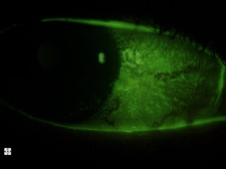
This is a pateint with rheumatoid arthritis with dry eye. You can see the fluorescent glow of the damaged cells on the surface in these photos. This patient already has punctal plugs but still has symptoms. We are starting her on a course of Restasis eye drops, but won't know for 3-6 months if they are working to reduce her symptoms and signs.

Wednesday, November 26, 2008

This patient has a "pterygium" on his left eye. This is scar tissue that develops from years of exposure to sunlight, dust and wind. It grows over the cornea with continued exposure and lack of sun protection. This patient's has remained this way for years.


This is the same patient's right lens. A slit of light is shined through the lens, so it's like looking at the lens in cross section, with the left side of it being the front of the lens and the right side being the back of the lens. You can see the layers in it. It's not a cataract unless these layers, usually the center, becomes cloudy. This is pretty normal lookingn.


Below is a photograph of the same patient's right eye with a nevus. The white dots are lipid deposits called "drusen" on the surface of the nevus. A nevus is like a mole in the eye. It just needs to be watched yearly to make sure it doesn't grow.


This is a picture of the same patient's lash ptosis. Most lashes come out or up. His point down. This condition does not bother the patients unless they actually curl into the cornea.


GLAUCOMA SUSPECT


This patient has intraocular pressures of 25mmHg in each eye with only moderate optic nerve cupping (as shown by the "donut hole" in the center of the optic nerve in each of these photos). His corneas were pretty normal thickness indicating that the pressure read clinically was pretty correct. He has some risk of glaucoma and will be tested with an automated visual field test and possibly a measurement of his nerve fiber layer thickness using an optical coherence tomographer.


The photo below is of pavingstone degeneration in the same patient's peripheral retina. This really does not affect the patient. I photographed it just because it's an interesting thing to see for patients.


This patient had "staining" along the superior cornea as shown here. Staining occurs when cells that have been sloughed off or damaged from any of a variety of causes. In this case no foreign body was found under the lid and no tear or rip was on the patient's contact lenses. That creates a clinical dilemma. However I did switch this patient's contact lens and the patient said it felt better. This picture is two days letter and the staining does look a little better. We'll keep monitoring.

Here you can see blood vessels growing into the cornea from long term use of contact lenses that don't breath enough oxygen. This patient has since been refit into Night and Day contact lenses.

Saturday, November 22, 2008

MORE CONTACT LENS-RELATED CORNEAL ULCERS


This is the left eye of a patient with an extremely busy lifestyle. This kind of patient will almost always wear their contact lenses all day and sleep in them often as well. There are only two brands of contact lenses that I feel comfortable fitting these patients in, Ciba Night and Day and Bausch and Lomb Purevision. The Purevision also comes in toric lenses for individuals with astigmatism.
Another interesting thing about this patient is that he originally went to an urgent care. Urgent cares, primary care providers, and emergency rooms typically give patients very mild antibiotic drops that are often not sufficient for the patient's eye condition. These kind of providers are limited in their ability to properly diagnose red eyes. This patient's problem persisted a week
beyond his visit to the urgent care. I gave him the appropriate drops that will clear the majority of the problem up within a few days.
Also this patient will need to be changed to Purevision Torics because of his astigmatism. Hopefully we'll find a pair that is comfortable for him physically and visually and we can decrease his risk of corneal ulcer.

Thursday, November 13, 2008

CORNEAL SCAR


This patient is now in his 50s, but suffered a scratch to the eye when he was a child. A surgeon attempted a procedure to laser off the front surface but it was unsuccessful since the scar was too deep. If this patient desires to see well again in this eye, they would have to go through the long and hard process of getting a corneal transplant, and then having a hard contact lens fit about a year later. In this case, it is best to protect the fellow eye with polycarbonate lenses, which are super strong.

Wednesday, November 12, 2008

SNOWFLAKE VITREORETINAL DEGENERATION


These are pictures of the peripheral retina in a patient's right eye and left eye. It demonstrates what we call Snowflake Vitreoretinal Degeneration. It most often found in near-sighted individuals and heredity plays a role. A small percentage of these patients develop retinal breaks that can be treated with laser to prevent retinal detachment. These patients should be followed up with a dilated examination on a yearly basis.

Thursday, October 30, 2008

RETENTION CYST


This is a fluid-filled, thin-walled cyst in the underside of the eyelid. It is benign, causes no harm, and usually does not need to be treated. It can be drained if it is bothersome to the patient.

LUPUS


Lupus is an autoimmune disease that can cause vasculitis as pictured here. It is inflammation in the blood vessels of the eye that can cause lack of oxygen getting to the retina. This results in loss of vision.

Monday, October 27, 2008

CONJUNCTIVAL GRANULOMA

This lesion under the eyelid caused this patient to literally cry blood from their eye. It is a painless, harmless, vascularized lesion that can be easily removed.

Thursday, October 23, 2008

GLAUCOMA


This is the optic nerve of one of my favorite patients. She has severe glaucoma with visual field loss. She is quite young and so must be treated very aggressively. Unfortunately she recently became sensitive to one of the most common glaucoma drops. But this can be managed with other options.
The crater in the middle of the optic nerve is called the optic "cup". The worse the glaucoma the deeper the cup. This is a very deep cup. The picture really doesn't do it justice because you would have to see it in 3-D.

CHRPE

This is CHRPE which stands for congenital hypertrophy of the retinal pigment epithelium. It is just a pigment spot on the inside wall of the eye. There is no risk of conversion to melanoma. It is just an interesting looking spot on the eye.

Tuesday, October 21, 2008

IRIS STRAND


This is an interesting finding in the same patient below. This is an iris strand that goes from one end of the iris, across the pupil, to the other end. It's sort of like a cobweb. It will not cause any harm and is not associated with any diseases of the eye as far as I know. It's just cool!

CORNEAL BLOOD VESSEL GROWTH


This is another case of a patient who overwears their contact lenses. They do not have a back up pair of glasses that they like. These blood vessels should not be growing here, and if nothing changes, eventually she may have problems with corneal ulcers and episodes of severe pain and light sensitivity. This patient got good glasses to wear. On a follow-up 6 months down the road, if the blood vessels aren't better, we'll go to the highest O2 breathing lens.

TILTED OPTIC NERVE


The bottom picture is of the optic nerve in the right eye of this young patient. The top one is the left eye. The one on top is tilted because the optic nerve enters the eyeball obliquely. This will cause no harm to the patient, except she see's 20/20 at distance with the right and 20/400 in the left. She is really near sighted in the left eye however but see's 20/20 up close in that eye. If her eyes don't change, she'll never need bifocals when she's past 45 years old.


PINGUECULA

This is a pinguecula. It is a type of scar tissue that develops from chonic exposure to UV light, dust, and wind. It is believed that it is mostly light from the side that causes this, so it is very important to have good sunglasses that wrap around and block the sides. We carry Kaenons and Rudy Projects as well as others, like Ray Bans, and Maui Jims. Kaenons are the best! Lightweight, really stylish, polarized, with anti-reflection on back side so you don't see your eyes.

Friday, October 17, 2008

HYPHEMA


This patient got hit by a pellet gun, causing a laceration (or abrasion) on the conjunctiva and a hyphema (bleeding in the anterior chamber).


Monday, October 13, 2008

OCULAR HYPERTENSION, GLAUCOMA SUSPICION

These are the right and left optic nerves of a patient with intraocular pressures of 25mmHg in each eye. Average pressure in a person's eye is 15mmHg. This patient has slightly thicker corneas than average, indicating that true intraocular pressure is a little bit lower than clinically measured intraocular pressure. The deeper and width of the cupping of the optic nerve, which corresponds a little beyond the whitened center in the photos below, the higher the risk or suspicion of glaucoma. This patient requires a visual field test and possibly some other tests to determine if the optic nerve is having any damage from the higher intraocular pressure. Those tests should be performed within 3 months for this patient.

Wednesday, October 8, 2008

MILD OR EARLY MACULAR DEGENERATION

There are a few small pigment spots in the "macular" area of this retina. There is no vision loss in this patient.

Monday, October 6, 2008

MARGINAL KERATITIS


Here you can see a subepithelial infiltrate which is a collection of white blood cells in response to inflammation. This is in a contact lens wearer who slept in non-extended wear contact lenses overnight. Most contact lens wearers with these problems do not have eyeglasses that are suitable for their lifestyle, therefore they depend on their contact lenses almost entirely. Patients tend to overwear their contact lenses when they don't have nice glasses that match their lifestyle.
In this case, it was the beginning of peripheral ulceration with multiple subepithelial infiltrates, therefore it was deemed inflammatory in nature. A combination antibiotic/anti-inflammatory was prescribed along with contact lens discontinuation during the healing period and close follow-up.