Search Eye Pictures

Monday, October 27, 2014

TREATED RETINAL HORSESHOE TEAR

The top arrow points to a laser scar. Laser scars surround the tear.  The bottom arrow points to the horseshoe tear.  This tear is about a year old and was located superiorly.  The patient had a fresh, untreated tear nasally in this same eye. 


Wednesday, October 22, 2014

WHITENED ARTERY

This is the retina on a woman in her mid 30's. If you click on the photo to enlarge it you can see the artery is not narrowed relative to the vein, but it is whitened.  The fellow eye additionally had cotton wool spots.  She has not been diagnosed with high cholesterol but her blood pressure was slightly elevated. 


ACQUIRED LASH CURLING

This was a woman who complained that her lashes in the corner were short for about a month.  Upon slit lamp examination it was revealed that she had sectoral lash curling. 


CONJUNCTIVAL ABRASION

This is an abrasion on the conjunctiva of a teenage girl who complained to her mother that her eye was irritated after an Arizona dust storm.  When I first looked at her the abrasion was not apparent but the area was very red.  Upon dye instillation and Wratten filter the abrasion revealed itself. 


BITOT'S SPOT

This is a small spot on the conjunctiva that does not wet well.  Many believe it to be associated with vitamin A deficiency. 


CORNEAL SCARRING FROM LONG-TERM SMALL DIAMETER RIGID GAS PERMEABLE CONTACT LENS WEAR

The patient below is in his 20's and has been wearing small diameter RGP's for about 15 years for keratoconus.  Both corneas show the same scarring.  He was referred to me for scleral lens fit and with scleral lenses we can get him to 20/25 in each eye while completely vaulting the entire cornea and providing excellent comfort!


Thursday, October 16, 2014

Wednesday, October 15, 2014

Wednesday, August 13, 2014

PERSISTENT HYALOID ARTERY

Persistent Hyaloid artery is usually in one eye only and occurs in 95% of premature infants.  It manifests as glial material from the optic nerve to the posterior surface of the lens, forming a Mittendorf's Dot.  It doesn't have symptoms for the patient and may sometimes be associated with optic disc coloboma or optic nerve hypoplasia.  


MACULAR CYST


Wednesday, August 6, 2014

BRANCH RETINAL VEIN OCCLUSION


This patient is in his early 50's. He reports not having HTN nor high cholesterol but he hasn't seen a doctor for a couple years.  His arteries are white and narrow, his blood pressure was 160/100, and he has significant corneal arcus. 


CORNEAL ABRASION FROM DOG SCRATCH


It's important here to check for Seidel Sign to make sure there was no penetrating injury.  Also is the dog current on it's shots? Has the patient had a current tetanus shot?


MILD EPITHELIAL INGROWTH POST-LASIK


Wednesday, July 23, 2014

ASTEROID HYALOSIS

Calcium soaps floating in the vitreous humor


INTERESTING CORNEAL FOREIGN BODY


Friday, July 11, 2014

IMPLANTABLE COLLAMER LENS

The Implantable Collamer Len (ICL) is a lens that is placed between the iris and the lens of the eye.  It is a good option for patients with between -3.00 and -6.00 of near-sightedness and the preferred refractive procedure for patients with -6.00 or worse.
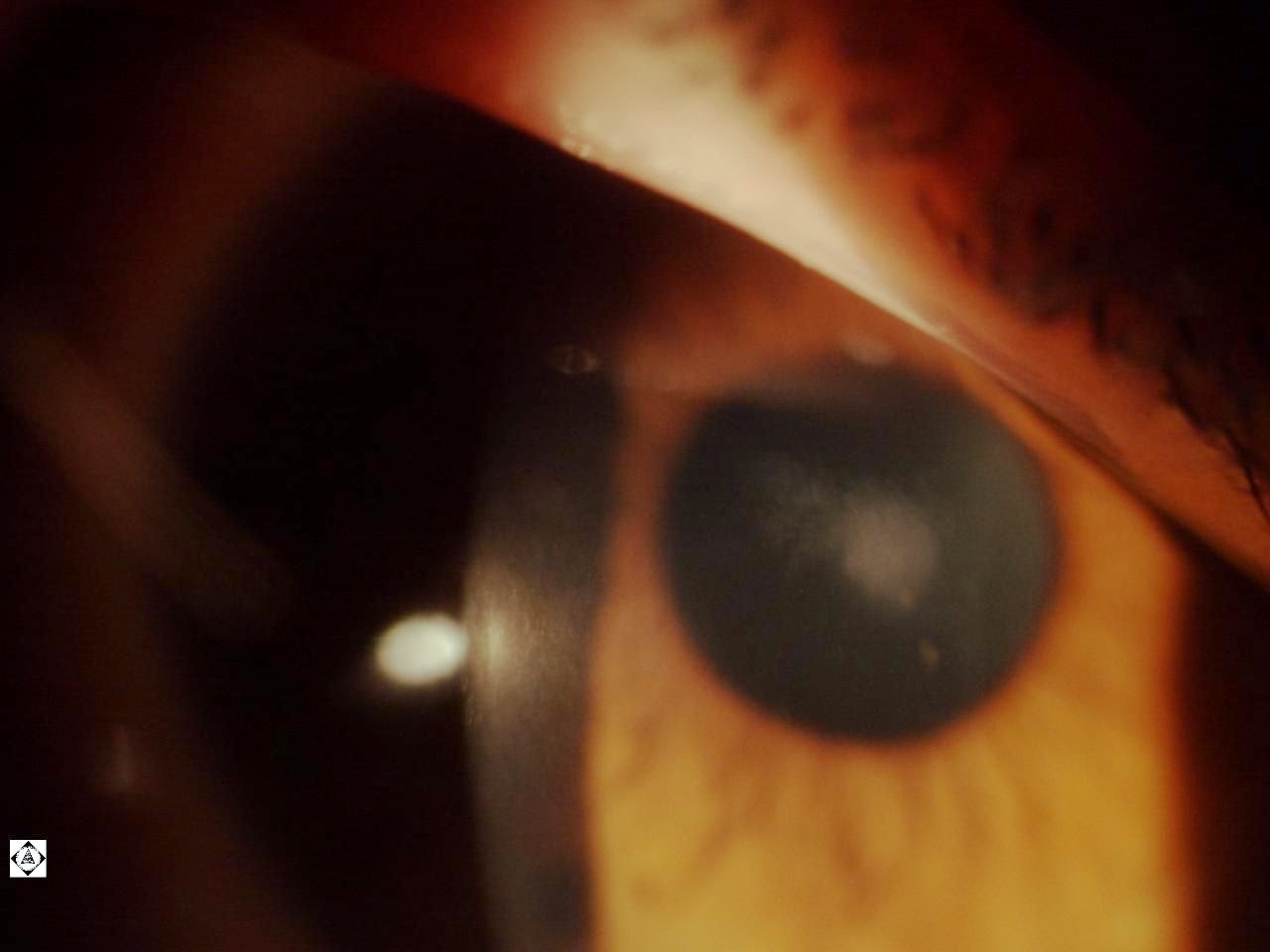
Thursday, June 26, 2014

TRAUMATIC IRIDODIALYSIS AND PROLAPSED IRIS

This gentleman poked his eye with the end of a sharp palm leaf while doing yardwork.  There is an iridodialysis from about 9:00 to 12:00, the anterior chamber is collapsed, and the iris is prolapsed through the full-thickness wound.  


PULVERULENT, COPPICK, OR EMBRYONIC CATARACT


Friday, June 13, 2014

REIS-BUCKLER'S' CORNEAL DYSTROPHY

Reis-Bucklers' corneal dystrophy is bilateral and is autosomal dominant with strong penetrance.

It presents in early childhood with the complaint of irritations and light sensitivity and then visual loss starts to occur as the cornea opacifies in the bowman's layer.  The corneal surface becomes very distorted and there is a honeycomb appearance. 

Hard lenses, especially scleral lenses, are beneficial for vision, even in moderate to advanced stages.  In severe cases corneal transplants can be performed, however recurrence of the dystrophy in the grafts is common.  Therapeutic Laser Keratectomy may also be performed. 

This photo is in a 32 year old male whose mother also had the condition. 


Tuesday, June 3, 2014

Wednesday, May 21, 2014

Monday, May 19, 2014

PALPEBRAL CONJUNCTIVAL FOREIGN BODY

A gentleman presented with extreme pain in his left eye.  It occurred while he was cutting prickly pear cactus and hour earlier. Slit lamp exam exam with fluorescein dye revealed the following. 


Vertical lines on the cornea indicate a foreign body on the palpebral conjunctiva under the upper eyelid.  


Lid eversion revealed an embedded cactus spine. 


Without the dye it is much more difficult to see the cactus spine. 


The spine was removed with a jewelers forceps. 


Further investigation revealed another spine in the eyelid near the meibomian gland openings.  No further spines were found on more extensive investigation. 

Thursday, May 8, 2014

PSEUDOEXFOLIATION


TRIPLE CATARACT

This patient has an anterior capsular, nuclear, and posterior subcapsular cataract. 


Wednesday, April 30, 2014

VORTEX KERATOPATHY

You can see the cornea verticullata from amioderone use.  Amioderone is a drug used in arythmia. 


SOLUTION SENSITIVITY

You can see the punctate epithelial erosions and unstable tear film on the cornea.  


ANTERIOR BASEMENT MEMBRANE DYSTROPHY (ABMD)

Anterior Basement Membrane Dystrophy (ABMD)


Monday, April 7, 2014

HERPES SIMPLEX VIRUS (HSV) KERATITIS

Note the pathogneumonic dendrite  in this HSV lesion


Friday, April 4, 2014

CORNEAL STAINING FROM SOLUTION SENSITIVITY

This is the cornea of a contact lens patient.  I was having great difficulty getting this patients better than 20/30 with refraction and discovered the punctate staining prior to checking pressures.  


Thursday, April 3, 2014

FUNDUS COLOBOMA

 This is the right eye of the patient and the left eye looks exactly the same.  This is often associated with developmental syndromes. In this case the patient has mild mental retardation and hearing loss.  However, the patient see's 20/20 in each eye.


Wednesday, March 26, 2014

CONJUNCTIVAL LITHIASIS (CONCRETIONS) AND CYSTS)


GUTTAE

Guttae are small excrecenses on the corneal endothelium. Also called guttata.  Often associated with Fuch's Corrneal Dystrophy. 


Tuesday, March 25, 2014

VOGT'S STRIA

You likely have to enlarge the image to see the vertical lines, called Vogt's Stria, on the back of the cornea.