Search Eye Pictures

Saturday, November 29, 2008

DRY EYE DAMAGE


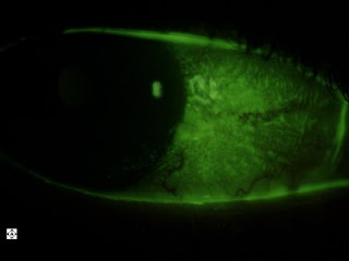
This is a pateint with rheumatoid arthritis with dry eye. You can see the fluorescent glow of the damaged cells on the surface in these photos. This patient already has punctal plugs but still has symptoms. We are starting her on a course of Restasis eye drops, but won't know for 3-6 months if they are working to reduce her symptoms and signs.

Wednesday, November 26, 2008

This patient has a "pterygium" on his left eye. This is scar tissue that develops from years of exposure to sunlight, dust and wind. It grows over the cornea with continued exposure and lack of sun protection. This patient's has remained this way for years.


This is the same patient's right lens. A slit of light is shined through the lens, so it's like looking at the lens in cross section, with the left side of it being the front of the lens and the right side being the back of the lens. You can see the layers in it. It's not a cataract unless these layers, usually the center, becomes cloudy. This is pretty normal lookingn.


Below is a photograph of the same patient's right eye with a nevus. The white dots are lipid deposits called "drusen" on the surface of the nevus. A nevus is like a mole in the eye. It just needs to be watched yearly to make sure it doesn't grow.


This is a picture of the same patient's lash ptosis. Most lashes come out or up. His point down. This condition does not bother the patients unless they actually curl into the cornea.


GLAUCOMA SUSPECT


This patient has intraocular pressures of 25mmHg in each eye with only moderate optic nerve cupping (as shown by the "donut hole" in the center of the optic nerve in each of these photos). His corneas were pretty normal thickness indicating that the pressure read clinically was pretty correct. He has some risk of glaucoma and will be tested with an automated visual field test and possibly a measurement of his nerve fiber layer thickness using an optical coherence tomographer.


The photo below is of pavingstone degeneration in the same patient's peripheral retina. This really does not affect the patient. I photographed it just because it's an interesting thing to see for patients.


This patient had "staining" along the superior cornea as shown here. Staining occurs when cells that have been sloughed off or damaged from any of a variety of causes. In this case no foreign body was found under the lid and no tear or rip was on the patient's contact lenses. That creates a clinical dilemma. However I did switch this patient's contact lens and the patient said it felt better. This picture is two days letter and the staining does look a little better. We'll keep monitoring.

Here you can see blood vessels growing into the cornea from long term use of contact lenses that don't breath enough oxygen. This patient has since been refit into Night and Day contact lenses.

Saturday, November 22, 2008

MORE CONTACT LENS-RELATED CORNEAL ULCERS


This is the left eye of a patient with an extremely busy lifestyle. This kind of patient will almost always wear their contact lenses all day and sleep in them often as well. There are only two brands of contact lenses that I feel comfortable fitting these patients in, Ciba Night and Day and Bausch and Lomb Purevision. The Purevision also comes in toric lenses for individuals with astigmatism.
Another interesting thing about this patient is that he originally went to an urgent care. Urgent cares, primary care providers, and emergency rooms typically give patients very mild antibiotic drops that are often not sufficient for the patient's eye condition. These kind of providers are limited in their ability to properly diagnose red eyes. This patient's problem persisted a week
beyond his visit to the urgent care. I gave him the appropriate drops that will clear the majority of the problem up within a few days.
Also this patient will need to be changed to Purevision Torics because of his astigmatism. Hopefully we'll find a pair that is comfortable for him physically and visually and we can decrease his risk of corneal ulcer.

Thursday, November 13, 2008

CORNEAL SCAR


This patient is now in his 50s, but suffered a scratch to the eye when he was a child. A surgeon attempted a procedure to laser off the front surface but it was unsuccessful since the scar was too deep. If this patient desires to see well again in this eye, they would have to go through the long and hard process of getting a corneal transplant, and then having a hard contact lens fit about a year later. In this case, it is best to protect the fellow eye with polycarbonate lenses, which are super strong.

Wednesday, November 12, 2008

SNOWFLAKE VITREORETINAL DEGENERATION


These are pictures of the peripheral retina in a patient's right eye and left eye. It demonstrates what we call Snowflake Vitreoretinal Degeneration. It most often found in near-sighted individuals and heredity plays a role. A small percentage of these patients develop retinal breaks that can be treated with laser to prevent retinal detachment. These patients should be followed up with a dilated examination on a yearly basis.