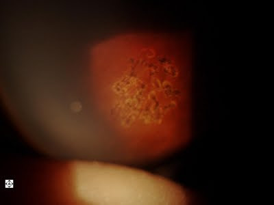
Otherwise known as Map, Dot, Fingerprint Dystrophy
Search Eye Pictures
Wednesday, December 14, 2011
Wednesday, December 7, 2011
Saturday, December 3, 2011
Palpebral conjunctival Papilloma vs. Granuloma
This is a lesion under the upper eyelid. The upper lid is everted. The top picture is taken in December 2011 and the bottom picture was taken in July 2011. The lesion appears less vascularized (less blood supply) curretnly, however it does appear like there are is a new nodule to the left and larger nodule to the right. With the evidence of growth, I believe that, although this is likely a benign lesion, it should be removed before it becomes much larger, so that it is easier to remove with less risk of complication than if we wait.
Tuesday, November 29, 2011
LARGE RETINAL HOLE
This is a large hole in the retina in a 20 year old patient. It needs fairly urgent attention before it becomes a retinal detachment. There is some fluid under it so part of the retina is already detached. We catch about 2 to 3 patients with retinal holes per week just on routine dilated eye exams. Without dilating, these breaks in the retina cannot be discovered. Therefore the people that go blind from retina detachments are often the ones with perfect vision who don't come in for exams regularly, because they don't get discovered until it's too late.
Friday, October 28, 2011
CORTICAL SPOKING CATARACTS
These are the kind of cataracts that grow from the outside of the lens, working it's way in. While it looks bad through the microscope, this patient is still able to achieve 20/20 vision, albeit not as sharp as we'd like. It causes glare problems and starbursts at night.
Friday, September 30, 2011
CORNEAL ABRASION
The patient in the photos below had her hard lens decenter off her eye and in the effort to remove it her cornea was abraded. So her vision is blurry, she's in pain, and she's light sensitive.
Tuesday, September 20, 2011
WHY SHOULD I REPLACE MY CONTACT LENSES MONTHLY
Below are photos of the underside of the lids of patients who overwear their contact lenses and don't replace them when they're supposed to. These lids have inflammatory bumps on them that make the contact lenses move, the vision fluctuate, and mucous come out of the eye. Nothing like mucous coming out of your eye when you're on a date!
GPC takes months to get rid of, and sometimes years. We have to use more expensive contact lenses thereafter and you have to use expensive drops and care solutions to try to fix the problem.
Bottom line? Please replace your contact lenses when you're supposed to. If you can't afford to replace your lenses monthly, don't get contact lenses. These are your eyes. Compromise elsewhere!
Monday, September 19, 2011
Tuesday, September 13, 2011
BITOT'S SPOT
A Bitot's Spot is an area on the white part of the eye that doesn't wet well and appears keratinized. It is often associated with vitamin A deficiency. It is fairly asymptomatic and patients are unaware they have it when it is discovered. Vitamin A deficiency happens mostly in children in developing countries, but we often see it in college kids for various reasons (including malnutrition and alcohol consumption). Bitot's Spot is often one of the first signs of Vitamin A deficiency. Advanced deficiency can lead to night blindness.
Monday, September 12, 2011
VITREOUS FLOATER
Our eyes are fluid-filled and you can often see floaters. The arrow points to a floater in this patient's eyes and the dark spot behind it is the shadow of the floater on the retina.
Friday, September 9, 2011
CORNEAL TRANSPLANT
This is what a cornea looks like after a cornea transplant. Best visual acuity is usually around 20/50 after a transplant with a significant amount of astigmatism. Transplants can become necessary if the cornea is scarred, or is shaped incorrectly, or has opacities or irregularities from genetic corneal diseases.
Friday, August 26, 2011
GLAUCOMA
One of the things we look at when we dilate the eye is the optic nerve cupping. It's the white area in the middle of the optic nerve in the photo below. The smaller the white area the less the risk for glaucoma. As pressure damages the optic nerve, the white area grows and your peripheral vision decreases until you're blind. The 2nd photo shows an eye with glaucoma. Note the absence of pink area inferiorly.
Thursday, August 25, 2011
Wednesday, August 24, 2011
COLD SORE ON THE EYE!
Herpes Simplex Virus resides in the nerve centers of about 95% of the population. It is responsible for cold sores on the lips. When it activates in the eye, it can cause discomfort, decreased vision, pain, light sensitivity, and watering. HSV can reactivate during periods of stress or with extreme exposure to sunlight.
When HSV activates on the eye, we treat it with antivirals and watch very carefully for scarring and secondary inflammatory damage. It can be very serious and even cause blindness if mismanaged.
Friday, July 29, 2011
Monday, July 25, 2011
Thursday, July 7, 2011
Friday, July 1, 2011
Friday, June 3, 2011
MYELINATED NERVE FIBERS
The bottom photograph is of a normal optic nerve. the top four demonstrate myelinated nerve fibers. Nerve fibers are usually insulated with myelin, that aids in transmission of nerve impulses. Usually this insulation ends as the fibers enter the eyeball. But occasionally, as is the case below, the insulation will continue, and you can see it as the white. These usually have no negative consequence to vision or the eye.
Wednesday, June 1, 2011
Friday, April 22, 2011
EYE DAMAGE FROM ACUVUE LENSES
Acuvue Oasys lenses are the most comfortable lenses on the market. Unfortunately, they cause more damage than any other lens I've seen. This occurs primarily because the edge of the contact lens steepens. In this case, you can see the damage from the edge hitting the eye. But because it is so comfortable, patients don't let us switch them to anything else, even if they KNOW there's a healthier alternative. These lenses are too dang comfortable! So my advice is to wear Acuvue Oasys lenses no more than 12 hours a day, 5 days a week. Those patients seem to do well with no damage. Whenever I see damage to the eyes, it's always from wearing the lenses longer than that. And don't wait to replace your lenses until the contact lenses are irritating and the eyes are already damaged. That's like waiting to do an oil change until the car smokes. Or waiting to add chlorine to your pool until it's filled with algae.
RETINAL HOLES
Every day people go blind from retinal detachments that could have been prevented if they had been caught as retinal holes. The retina is so thin that holes can develop in them spontaneously. Then fluid gets under the holds and detaches the retina, and where the retina detaches, you can't see. This is a picture of a patient who gets the eyes checked every year. Never have retinal holes been found before on this patient. It is important to dilate the eyes on your routine eye exam. It is also important that people have eye exams, even if they don't need glasses or contact lenses.
Click on the pictures below to enlarge
Tuesday, April 5, 2011
ASTEROID HYALOSIS
Asteroid hyalosis is calcium floating inside the fluid of the eye. It is generally not noticed by patients and does not need to be treated.
Wednesday, March 23, 2011
BUMP ON THE EYE
A chalazion is a bump on the lid that is non-tender and non-infectious. It arises because of stagnation of secretions from a clogged meibomian glands. It is initially treated with aggressive hot compresses and steroid drops. Sometimes steroid is injected into it. Often, if these measures don't work, it has to be removed surgically. This is not painful as the surgeon numbs the eye.
Tuesday, March 22, 2011
ONE REASON YOU SHOULD WEAR SUNGLASSES
A person who spends a lot of time in the sun should definitely use sunglasses. One of the things that can happen is that scar tissue extends onto the cornea because of UV light exposure. This scar tissue will continue to grow until it covers the whole eye. It can be surgically removed before it does so. This lesion is called a pterygium (pronounced "tuh-RIG-ee-um")
Saturday, March 19, 2011
Crusts in The Eyes in the Mornings
The picture below is the eyelid of an 18 year old boy. He has crusts at the base of his lashes. These crusts are produced from bacteria in the eyelash follicles. This condition is chronic and called blepharitis. It can cause a foreign body sensation, redness, and irritation of the eyes in the mornings. It can be controlled with lid scrubs and occasional antibiotics, but is never eradicated completely.
Friday, March 18, 2011
Red and Irritated Eyes
Sometimes the eyes feel are red in the mornings and they feel irritated. In the picture below the patient has a lot of debris in their tear film, the eyes are a little red, and there are clogged glands on the lid margin. This is called meibomianitis. This is a chronic condition that comes from overpopulation of the normal bacteria in the eyelid because of glands having a tendency to clog. Treatment ranges from hot compresses (rubbing the eyes well with a very hot cloth a few times a day) to doxycycline pills on a long term basis. Omega-3 fatty acid supplementation may also help. When the eye becomes very inflammed to where it causes light sensitivity and pain, we have to use combination antibiotic/steroid drops.
Thursday, March 17, 2011
Dry Eyes
The eye below is dry. Instead of a nice smooth tear film it is broken up quickly. You can also see some damage to some corneal surface cells.
Thursday, March 10, 2011
"MY CATARACT'S COMING BACK!"
About 35% of patients that have cataract surgery can develop a film on the implant during the first year after surgery. Sometimes this film can be so bad that it decreases the vision and the patient feels like the cataract is back. Patients feel like they're looking through a film.
These cloudy capsules can be easily cleared with a painless laser procedure called a YAG Capsulotomy. Usually the film never grows back again.
Subscribe to:
Comments (Atom)


