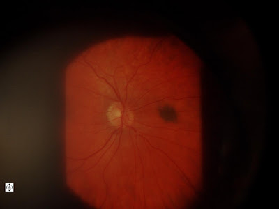
The photo below shows a brown spot next to the optic nerve in this young patient. The brown spot is a choroidal nevus that corresponds to pigmented spots on the skin that we consider normal. It has potential for growth, even to a malignant melanoma. So we document it with photos and watch it closely. At Sonoran Deseert Eye Center we have the capability to take these photos, rather than have to send you to a specialist to perform these photos.
Search Eye Pictures
Saturday, February 26, 2011
Thursday, February 24, 2011
More Drusen
The picture below are more drusen deposits along the superior arcade. These drusen are in younger patients and are likely harmless.
Wednesday, February 23, 2011
Fluffy White Dots in the Eye
The picture below shows fluffy white dots in the macula area of the eye's retina. The macula serves the center of the vision. This patient came in for a routine eye exam and see's 20/20. These dots are called drusen, and are deposits of waste material that the bloodstream doesn't absorb easily. Since the macula is the area of the retina that has the most metabolic activity, because it serves our central vision, it tends to get more accumulation of drusen. This drusen accumulation causes eventual damage to the macula resulting in vision loss. This vision loss is called macular degeneration. Because we see that she has macular degeneration, we were able to get her on the proper multi-vitamins and diet to help decrease risk of progression.
Tuesday, February 22, 2011
Clogged Blood Vessel in the Eye
The eye is often the first place that cardiovascular disease shows up. On a routine examination, an embolus was seen in this eye below. This alerts us to other cardiovascular problems and likely blockage in larger blood vessels, such as the carotid arteries. Eye exams are truly about "seeing the signs" warning us of what's coming up.
Monday, February 21, 2011
Seeing double in one eye
The picture below is a patient who had cataract surgery and an implant put in their eye. Usually cataract surgery goes smoothly with no complication. In this case, the patient's intraocular implant moved a little so that the edge of it appears in the patient's visual axis. We call this subluxation or a subluxated implant. It can be fixed by the surgeon.
Friday, February 18, 2011
My Eye is Red
Redness in the eyes can be caused by infection, inflammation, high blood pressure, allergy, dry eye, foreign body, growths, and others. It can be difficult to determine the cause of inflammation treating for one thing may not be appropriate for another.
This patient woke up with redness on the top part of the eye. It is mildly uncomfortable. There is actually a nodule or bump and there are no abrasions on the surface. There is no mucous coming out of the eye. This is an episcleritis, specifically nodular episcleritis. It is treated with topical steroids and/or oral non-steroidal anti-inflammatories. It is a non-infectious inflammation, although I have seen herpes simplex virus masquerade as episcleritis. That is why is it very important for a doctor to follow a patient using topical steroids very very closely, as steroids will make herpes simplex much worse. Doctors must feel comfortable managing this or they should refer.
Thursday, February 17, 2011
Tuesday, February 15, 2011
Birth defect in eye
The picture on top is a normal looking optic nerve. The one on the bottom is one that has a coloboma. This is a birth defect that occurs because of inadequate closure of the fetal fissure. Fortunately, this patient has normal vision.
Monday, February 14, 2011
Flakes in The Eye
You can see a white, flaky, ring on the surface of the lens in the picture below. In some people of Scandinavian descent, a flaky material will form on the front of the lens. Then as the pupil constricts it pushes this flaky material into this ring. In some situations this flaky substance can actually block fluid drainage in the eye, causing the pressure to rise and increasing the risk of glaucoma. This flaky material is called pseudoexfoliation and patients with this condition have to be watched closely for glaucoma.
Thursday, February 10, 2011
Irritated Eyes
The picture below shows the cause of irritated eyes. I dyed the tears and the picture shows how the tears break up. There are also some damaged cells at the bottom of the cornea. These are signs of dry eye, which can be treated.
Wednesday, February 9, 2011
I can't see!
The virus that causes cold sores on the lips can also cause "cold sores" on the eye. The picture below is not an ulcerative herpes simplex lesion but is an inflammation that occurs because of the virus, without the primary infection . After initial infection the virus harbors itself in the nerves and remains inactive. During periods of stress, the virus is triggered and activated and causes problems in the eye.
In the case below the patient cleared with a combination of anti-virals and steroid drops. This condition has to be managed very very carefully.
Tuesday, February 8, 2011
There's a Bump on My Eye!
The bump in the corner of the eye is called a pinguecula. It is scar tissue from years of exposure to sun, dust and wind. It usually has a yellowish color to it. It can become red and inflammed sometimes. It can also feel dry on top of it causing a sandy foreign body sensation in the eye. An inflammed pinguecula can be treated with drops. It is recommended you use sunglasses outside.
Saturday, February 5, 2011
EYELASH STUCK IN MEIBOMIAN GLAND
This obviously irritates and scratches the eye and won't come out with rubbing the eyes. It seems very unlikely a loose lash would ever fall into a meibomian gland but it is surprisingly a common occurrence. It is really easy to remove with a jeweler's forceps in the microscope.
Friday, February 4, 2011
VITREORETINAL TUFT
A tuft on the retina is a small elevation where the bag of gel that fills the eye is pulling on a focal part of the retina. However there is no tear in the retina. It is higher risk for a tear and the patient should know to come in if they see flashes of light, spiderwebs in their vision. or a shadow or curtain coming over their vision. Otherwise we should monitor it yearly.
Cornea Scar
There is a scar on this patients' cornea, presumably from a scratch. However, this patient doesn't remember scratching the eye.
Whitened arterioles
The arterioles in the retina on the top picture are normal. The bottom arterioles appear to have some whitening. In someone in their 30's we become a little concerned about cholesterol levels.
Thursday, February 3, 2011
The effects of 2 week senofilcon—A silicone hydrogel contact lens daily wear on tear functions and ocular surface health status
The effects of 2 week senofilcon—A silicone hydrogel contact lens daily wear on tear functions and ocular surface health status
Objective tests for dry eye appear worse with silocone hydrogel contact lens use.
A Randomized Trial of Brimonidine Versus Timolol in Preserving Visual Function: Results From the Low-pressure Glaucoma Treatment Study
A Randomized Trial of Brimonidine Versus Timolol in Preserving Visual Function: Results From the Low-pressure Glaucoma Treatment Study
Brimonodine is better than timolol at preserving visual function.
Vitamin A Deficiency
Vitamin A deficiency can occur in aloholics and can cause dry, keratinized spots on the surface of the eye. These spots are called Bitot's Spots.
Wednesday, February 2, 2011
WHY IS THIS EYE TEARING?
There is a canal that goes from our eyes to our nose. Every time we blink, tears get pumped into the canal, so that they don't flow out onto our cheek.
In the case below, the patient was complaining of excessive tearing. In the corner of his eye, on the lower lid, you can see the opening of the canal. You may also see that the opening is not flush against the eyeball. The excess tears have no way of getting down into the canal.
In this case, if the excessive tearing bothers the patient enough, a surgical procedure can be performed to bring the opening of the canal back against the eyeball where the tears are.
This condition, called ectropion, happens mostly as we age and the lid becomes lax. It just falls away from the eye. It can also occur in the case of injury or growing lid lesions.
Tearing in the eye can sometimes be very difficult to diagnose. It can occur from dry eyes, clogged tear drainage, allergy, viral infection, or other inflammations.
For more information, contact us at Sonoran Desert Eye Center 480-812-2211, or visit our web page at www.SonoranDesertEye.com
Tuesday, February 1, 2011
Choroidal Folds
The picture below is of the retina on the back of the eye. The arrow points to a horizontal line. If you click on the picture you can see the lines more distinctly. These lines are choroidal folds, or folds in a layer of the eye. These folds occur when there is something behind the eyeball pushing forward on the eye. This can be an orbital tumor. The patient may be otherwise asymptomatic.
Subscribe to:
Comments (Atom)


