Search Eye Pictures

Monday, January 31, 2011

Routine Eye Exam Reveals Increased Brain Pressure

Papilledema is a swollen optic nerve head in the eye.  The optic nerve connects the eyeball to the brain.  If there is pressure in the brain, it will cause the optic nerve to swell where it enters the eye.  The bottom picture below is the normal optic nerve and the top one is a swollen optic nerve head.   Note the indistinct margins of the optic nerve on top.  

Increased pressure in the brain can occur with or without a space-occupying brain lesion.  It is important to rule out any kind of space-occupying lesion such as a brain tumor.  This patient had no symptoms.  She was just coming in for a routine eye examination.  Sometimes patients with papilledema complain of headaches and ringing in the ears. 


Saturday, January 29, 2011

Lens Zonules


This is a patient with an iridodialysis, which is a section of the iris that has a hole in it near it's insertion.  Through that hole you can see the edge of the lens and the zonules that support the lens.  The zonules are thousands of thin strings that surround the lens and connect it to the ciliary body, the muscle that is responsible for focusing.  It's interesting that the lens of the eye is actually suspended in the by these cords.  When the muscle contracts for focusing at near, the zonules relax, allowing the lens to fatten in the middle, changing it's refractive power.  We don't often get to see the zonules.  If you click on the picture to enlarge it you may see the zonules more. 

Friday, January 28, 2011

Conjunctival Nevus

A nevus is a cluster of pigmented cells and just needs to be photographed and watched. 

TORTUOUS RETINAL VESSELS

Wednesday, January 26, 2011

GLAUCOMA SHUNT


Glaucoma occurs when high pressure in the eye damages the optic nerve, causing gradual loss of vision.  It is normally treated with eyedrops.  If eyedrops aren't sufficient then there are laser procedures that can help the fluid in the eye drain faster, decreasing the pressure in the eye.  If that does not lower the pressure enough, there is a bypass procedure that creates an alternate path for fluid to leave the eye.  

If drops and surgery is not enough to lower pressure sufficiently, a shunt can be placed in the eye.  The shunt is placed at the top of the eye and a tube extends from the shunt into the anterior chamber of the eye.  This procedure is reserved for those with significant, uncontrolled glaucoma and is considered a last-ditch effort to saving the eye and whatever vision is left. 

Tuesday, January 25, 2011

DRY EYE


The picture below was taken with dye in the eye to stain the tears.  You can see that the tear layer is very thin in places.  This causes irritation on people's eyes.  You can also see the lower part of the cornea is stained in spots.  This is damage that occurs to the surface cells because of dryness. 

Dry eye is managed first with different types of artificial tears, then with plugs or Restasis eye drops. 

Monday, January 24, 2011

Cataract


The picture below shows what a cataract looks like through the eye doctor's microscope.  A slit of light coming from the side shows a cross-section of the lens, where you can see a cloudy center.  Of course that cloudy center causes blurred vision and glare.  Cataract surgery is very successful with few complications and often people are less dependent on glasses after cataract surgery. 

Friday, January 21, 2011

Why not wear a two week lens for two months?


During this rough economy everyone is trying to save a buck.   What's wrong with making a 2-week disposable contact lens last for a month or two?

Well there are certainly a lot of people who do this without problems.  But when there are problems, it is always these people.  

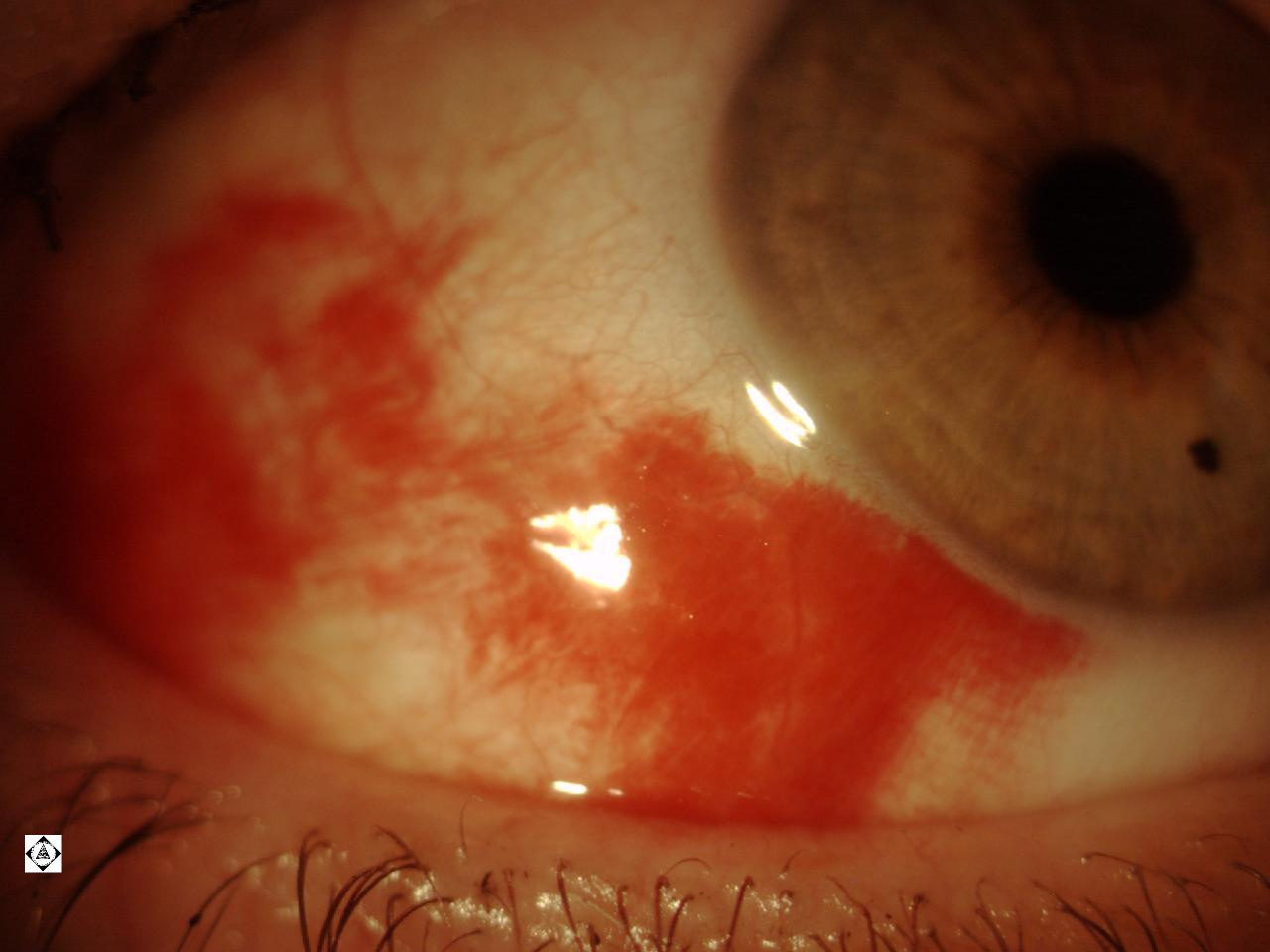
What are the problems that develop?  On the picture below you can see protein deposits from the tear film that develop on the surface of the contact lens.  You can also see blood vessels growing in to the cornea, demonstrating that the cornea is starved for oxygen from contact lens overwear. 


These deposits not only make the lens perform less, both in comfort and in vision, but they cause an inflammatory reaction in the eye.  Inflammatory bumps begin to form under the eyelid (see picture below).  The eyes begin to feel itcy and irritated.  The bumps pull on the contact lenses causing them to move more, which causes greater discomfort and fluctuating vision. 


Not only do protein deposits form on the contact lenses, but excretions from bacteria in the glands of the eyelids also begin to concentrate on the contact lenses.  The eye will have an acute reaction to this, causing ulceration, pain, light-sensitivity, and contact lens intolerance.   This occurs without warning. Both these conditions take time to treat and sometimes result in inability to wear contact lenses again in the future. 


These situations are very common in the office.  The bottom line is, follow the directions of your eyecare provider.  There's a reason we want you to replace your contact lenses.  Your eyes are worth it!  

Thursday, January 20, 2011

Subconjunctival Hemorrhage

So you wake up and you see your eye is really red like this picture below.  It scares you to death!  The eye doesn't hurt and it doesn't seem to be affecting the vision.  So what's the problem?

Although this looks awful, it is one of the most harmless things that can happen to your eye.  It is what we call a subconjunctival hemorrhage.  The blood vessels on the white part of the eye can be very brittle and may burst with the slightest pressure change.  So it can happen when a person coughs, sneezes, bends over to pick something up, lifts something heavy, or hangs upside down.  It can also happen spontaneously.

It clears up within days to weeks without any insult to the eye. If it recurs, then we have to consider blood tests to rule out decreased platelet count that can occur in people with anemia and other disorders.  If there's any pain, vision decrease, swelling, or discharge, then it's more than just a bleed and should be seen. 

Wednesday, January 19, 2011

HABITUAL PULLING OF EYELASHES

This is a picture of a patient who has the nervous habit of pulling on their eyelashes.  As you can tell, many of the eyelashes are missing.  This is called trichotillomania.  It can often be overcome with behavior feedback.