

Thursday, December 26, 2019
Tuesday, December 17, 2019

Tuesday, December 10, 2019
RETINOSCHISIS
Look closely and you will see a retinoschisis in the right eye near the very bottom and the left eye around two o'clock (the right eye is the image on the left). A retinoschisis is a detachment of the retina from itself. A retinal detachment is when all of the nine layers of the retina detach from the underlying choroid. Retinoschisis does not normally need to be treated and rarely involves the posterior pole. Close monitoring is warranted.

Monday, December 2, 2019
RETINOSCHISIS
Below is a picture of a patient with a retinoschisis with an outer layer break in the retina. The lower arrow points to the break and the higher arrow points to the edge of the schisis. A schisis is a separation of the retina from itself. The edge of the schisis is usually smooth and in a circular pattern as shown here.


Wednesday, November 20, 2019

Friday, November 8, 2019

Thursday, October 31, 2019

Tuesday, October 29, 2019


Friday, October 25, 2019
ACUTE ZONAL OCCULT OUTER RETINOPATHY (AZOOR)
AZOOR is a rare retinal inflammatory disorder that can occur in young myopic females. Often it occurs in only one eye. It effects most often the temporal retina and spares the macula. It results in the type of scarring that you see below. This patient was myopic with normal central visual acuity.

Tuesday, August 6, 2019
ABMD Dots
These are likely the "dots" that we sometimes see in anterior basement membrane dystrophy.


Tuesday, July 30, 2019

Wednesday, July 10, 2019
BAND KERATOPATHY

Band keratopathy affects the cornea, the front clear window of the eye. Band keratopathy is a calcium deposit that appears grayish with holes in the middle, like swiss cheese. It starts from the 9:00 and 3:00 edge of the cornea and works it's way toward the center. Rarely it can encroach on the visual axis, and in these cases it can be removed by a process called chelation.
Band keratopathy can be caused by anything that increases calcium in the body. Some of these are as follows: kidney disease, excessive vitamin D, high thyroid levels, sarcoidosis, lupus and Paget’s disease. Also there is calcium in the tears.
Band keratopathy can be associated with several other eye conditions including severe glaucoma, chronic uveitis, corneal dystrophies, and phtisis bulbi (shrunken eye from severe loss of vision).
In most situation band keratopathy has no symptoms. Occasionally it can cause redness, irritation, and vision loss, depending on how advanced it is.
Chelation is a process that uses a special chemical and an excimer laser to remove the deposits.
Wednesday, June 19, 2019
DIABETIC RETINOPATHY
Diabetic retinopathy has been the number one cause of preventable blindness in the United States. Fortunately treatments have improved through the years.
The pictures below show patients with moderate non-proliferative diabetic retinopathy. There are various types of hemorrhages. Cotton wool spots are areas of focal oxygen deprivation. microaneurysms are outpouchings of capillaries that can leak. Exudates are the leakage of blood serum from capillaries.


If the oxygen deprivation is severe enough, new blood vessels (neovascularization) will begin to form in the attempt to reperfuse the retinal tissue. However, these new blood vessels are very leaky and certain contents from the blood destroy the retina, thereby causing severe vision loss. When new blood vessels grow, retina specialists often will perform pan-retinal photocoagulation, or PRP. PRP selectively destroys much of the peripheral retina, thereby decreasing the oxygen deman in the eye. This decreased oxygen demand allows the most important central part of the eye to receive enough oxygen to prevent neovascularization. The photo below shows the laser scars that result in the peripheral retina from PRP.

Thursday, June 13, 2019
TREATED RETINAL TEAR
This picture shows laser scars around the tear in the retina that would have ended up as a detached retina with accompanying permanent loss of vision.


ASTEROID HYALOSIS
Asteroid hyalosis is small white opacities that float in the eye. They uysually occur in one eye more than the other, as is the case with the picture below. It occurs in humans, dogs, cats, and horses. Ocular asteroids are different from the more common typical vitreous floaters. The cause of asteroid hyalosis is unknown, but it has been associated with diabetes , high blood pressure, and high cholesterol. The asteroid bodies are made up of calcium and phospholipids. While asteroid hyalosis is quite impressive, surprisingly most patients never notice them in their vision. They cause no problems, but may interfere with visualization of the retina during eye exams. Treatment of asteroid hyalosis is unnecessary unless better visualization of the retina is needed, such as in cases of diabetic retinopathy.


Wednesday, June 12, 2019
TREATED RETINAL HOLE
The image above is the treated hole with laser. The image below is the original hole. 


Friday, June 7, 2019
BRANCH RETINAL ARTERY OCCLUSION

Branch retinal artery occlusion in a 54 year old male. It caused retinal neovascularization which then lead to a vitreous hemorrhage.
Tuesday, June 4, 2019
BEAR TRACKS
The image below displays higher concentrations of pigment in the retinal pigment epithelium layer. They have the appearance of "bear tracks" and are harmless.


SCUBA DIVING MASK SQUEEZE
A patient presented after a scuba trip with the following presentation:

Failure to equalize the mask on a dive descent will result in suctioning of the mask, which will cause blood vessels to enlarge and leak, resulting in the bleeding and swelling. This is called "mask squeeze". The bleeding is harmless and will resolve on it's own within weeks.

Failure to equalize the mask on a dive descent will result in suctioning of the mask, which will cause blood vessels to enlarge and leak, resulting in the bleeding and swelling. This is called "mask squeeze". The bleeding is harmless and will resolve on it's own within weeks.
Friday, May 24, 2019
MYELINATED NERVE FIBERS

CORNEAL ULCER
A 19 year old female presented with the chief complaint of a painful, light sensitive right eye. She normally wear a Biofinity toric contact lens in the right eye and a Biofinity spherical lens in the left eye about 16 hours per day. A single, mid-peripheral corneal ulcer was responsible for her symptoms. The cornea is the only structure in the body that gets it's oxygen from the air. When you wear a contact lens, it reduces the oxygen getting to the corneal surface. The cornea can tolerate it up to 12 hours per day in general, but beyond this the cornea becomes hypoxic, or oxygen deprived. Bacteria love this hot, moist, anaerobic environment and the cornea suddenly becomes a petri dish for bacteria to grow and eat away at the cornea. Sometimes these ulcers can occur in the very center of the vision and cause permanent scarring that can permanently affect the vision. Some ulcers can be so bad as to cause a complete loss of the cornea and eye. Symptoms begin suddenly, so it is extremely important that patients wear properly fitted contact lenses according to the regimen prescribed by their doctor.
 |
| Corneal ulcer with white blood cells underlying |
 |
| Corneal ulcer with fluorescein stain |
Tuesday, April 23, 2019
HERPES SIMPLEX VIRUS DISCIFORM KERATITIS
A college-aged patient came in just before final exams complaining of blurred vision in the right eye. The eye was a little red and the vision was down to 20/40. The cornea was swollen with subepithelial and endothelial infiltrates as shown below. The patient has HSV disciform keratitis which requires topical steroids and oral anti-virals. Recalcitrant or recurrent disease sometimes requires endothelial surgery to rehabilitate the cornea.
Saturday, April 20, 2019
EPIBLEPHARON
Epiblepharon occurs when the lashes turn in even though the eyelid position is normal. It's typically seen in children and young adults of Asian descent. Affected patients have a congenital horizontal fold of skin near the lower eyelid that pushes the eyelashes toward the eyeball. In this case, you can see the lashes scratching the surface of the cornea.

Wednesday, March 13, 2019
ASTEROID HYALOSIS
The amazing thing about asteroid hyalosis is that the patient doesn't have decreased vision or see floaters!


Friday, March 8, 2019

Thursday, January 31, 2019

Internal Hordeolum
 Patient entered with a complaint of her right lower eyelid being painful, swollen, and red. Main differentials based on location of the event included preseptal cellulitis, lacrimal canaliculitis, and hordeolum. Flipping of the lower eyelid revealed an internal hordeolum within a meibomian gland adjacent to the lacrimal canaliculus.
 Wednesday, January 9, 2019
SEROUS RETINAL DETACHMENT FROM POSSIBLE CHOROIDAL NEOVASCULAR NET
In the picture below, the photo on the right is from March 2018. The one on the left is from January 2019. Visual acuity is unaffected. There is an unchanged chorioretinal scar near the macula. But where the arrows point, there is more subretinal fluid in the current photo.

Fundus Autofluorescence below demonstrates an increasing amount of hyperautofluorescence over the area of concern.


A cross-line OCT focused on the area of concern demonstrates a focal sub-RPE lesion pushing inward, a serous retinal detachment surrounding it, and a thin line demonstrating the vitreous membrane.

Tuesday, January 8, 2019
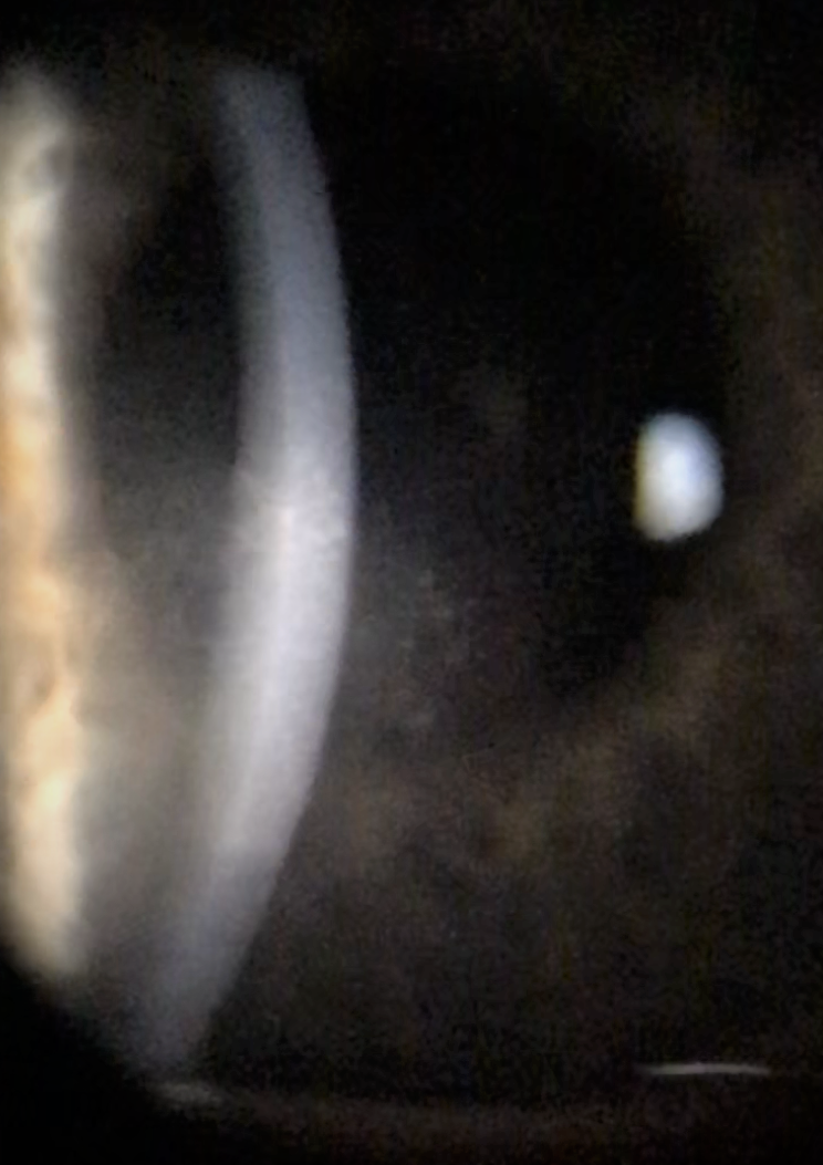
WEISS' RING FROM POSTERIOR VITREOUS DETACHMENT

The arrow shows a Weiss' Ring. There is a clear vitreous membrane that detaches off the retina at some point in most people's lives. A Weiss' Ring is where that membrane was attached to the optic nerve.
Subscribe to:
Posts (Atom)