The images below were shot with our Optomap Technology. The top images are the right and left eye. You can see optic nerve drusen below the optic nerve in the left eye. These drusen are confirmed with hyperfluorescence at the optic nerve with autofluorescence.
Search Eye Pictures
Friday, August 25, 2017
Wednesday, June 21, 2017
OPTIC NERVE HYPOPLASIA
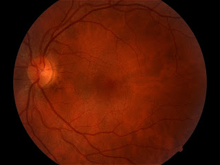
These are photos of the posterior pole of a 31 year old female with best visual acuity of 20/50 in the right eye and 20/15 in the left.
The left fundus looks normal.
The right optic nerve has white, fibrous tissue attached to it which I'm calling fetal tissue, even though it's unusual. There is some macular epiretinal wrinkling in a pattern that appears to be more tractional than puckering, similar perhaps to that seen in cases of toxocara. But the other interesting point is that the optic nerve also appears to be underdeveloped, and the blood vessels emanate more from the rim than from the center, suggestive of optic nerve hypoplasia.
Thursday, May 4, 2017
SCLERAL LENS COMPRESSION
The patient in the photo below presented with the complaint of red eyes while wearing his scleral contact lens. A scleral contact lens is a rigid gas permeable lens that vaults the cornea and is supposed to land softly on the conjunctiva. If it lands too steep it will compress blood vessels causing congestion and inflammation. This patient was refit successfully into a scleral lens that had a much softer landing and the redness subsided.
VITREORETINAL TRACTION
The above picture is an optical coherence tomography of the macular area of a patient with vitreomacular traction. You can see the clear vitreous membrane detached from most of the macular area but tightly adherent to the inner limiting membrane at the fovea. It pulls ont he membrane causing a small underlying cystic space.
Subscribe to:
Posts (Atom)


