Search Eye Pictures

Wednesday, August 4, 2010

CONTACT LENS OVERWEAR


This patient overwears their contact lenses and you can see blood vessels growing into the cornea. This is a common problem and people are surprised to see that contact lenses are causing this kind of damage to their eyes.

NUCLEAR CATARACT

The right lens shows barely any cataract at all.

The left lens shows a clouding which changed the patient's eyeglass prescription rapidly and will eventually cloud the vision to the point where he needs cataract surgery.

Friday, July 23, 2010

WASH YOUR HANDS BEFORE PUTTING IN CONTACT LENSES!!!!!

This is the case of a young lady who may not have washed her hands to put her contact lenses in after she had used some facial cream. The cream may be alkaline which is NOT good for the tissues of the eye. Alkaline substances tend to start damaging the cornea and working it's way deeper and deeper into the tissue.

On her way to work she started seeing hazy, but it didn't hurt. She came into the office and she really didn't look that bad, just a little damage to some surface cells of her cornea and a lot of corneal swelling. I had her irrigate her eye and use a lot of artificial tears the rest of the day to wash out everything and lubricate the eye. She called me last night in pain and I prescribed an antiobiotic/steroid combination eyedrop to calm down the inflammation and prevent bacterial infection.

Today she feels a lot better, and although her corneal swelling is a lot better, she has a lot of damage to the epithelial cells and her vision is worse. You can see that damage in the pictures.

Steroids can delay wound healing so, after consulting with my favorite corneal specialist, who concurs with me, I am having her use antibiotic drops only.

She will heal and I will watch her closely to make sure she heals with minimal or no scarring. However, the lesson is learned to WASH YOUR HANDS before putting contact lenses in. Washing hands is not just to prevent infection, but to wash off anything that the hands touched that could be toxic to the cornea.

Below is right eye yesterday.


Below is right eye today, showing the damaged epithelium cells.


This photo below is the left eye yesterday showing mild damage to surface cells.


The picture below is the left eye today, showing two heavily damaged areas of epithelium.


Below is the OS lesion using white light.


Tuesday, July 13, 2010

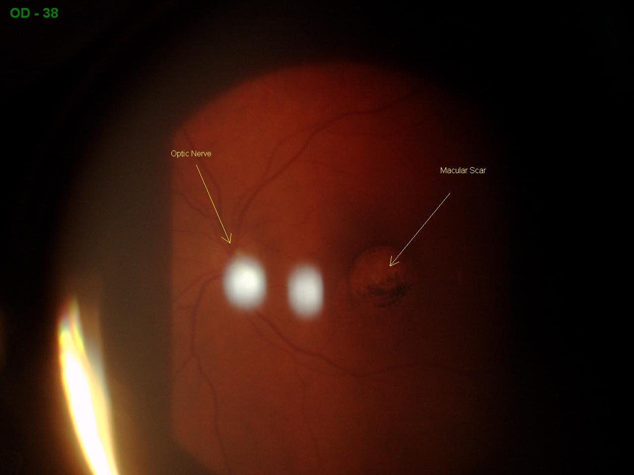
MACULAR SCAR

This is a scar from childbirth near the macula. The macula is that portion of the retina that serves the center of our vision. Because this scar is so close to the macula, this patient's best vision is 20/40. He has to eccentrically view, which means that he has to look off to the side a little to see something. Luckily his left eye is perfect 20/20.


Wednesday, June 30, 2010

SNAIL-TRACK DEGENERATION

This area of white is on the retina in the far periphery. It can only be seen when the eye is dilated. This occurs often as an inherited trait in near-sighted people. People that have this are more susceptible to developing retinal holes and detachments. This picture is of a young patient with the condition. It was discovered without any symptoms on routine dilation. She was educated to watch for flashes of light, floaters, or a curtain coming over her vision. We will follow up with her on a yearly basis.

Tuesday, June 29, 2010

METAL IN THE EYE


This mechanic's eye started bothering him on Tuesday. He thought it would go away on it's own but everyday it got worse until I saw him in my office on Saturday. He had metal embedded on his cornea. Removal was quick, will leave no scar, and provided instantaneous relief.

AIR HOCKEY INJURY


This patient was watching an air hockey match when the puck flew off the table and hit his eyelid. It lacerated his lid, which will heal rapidly. I dilated the eye to make sure there was no serious injury to the retina and found this area of whitening on the retina corresponding to the location he was hit.

The whitened area of the retina is what we call Commotio Retinae. It is caused by swelling in the retina from trauma. It resolves in about 6 weeks without any consequence to vision or health of the eye.