Search Eye Pictures

Friday, June 7, 2019

BRANCH RETINAL ARTERY OCCLUSION

Branch retinal artery occlusion in a 54 year old male.  It caused retinal neovascularization which then lead to a vitreous hemorrhage.  

Tuesday, June 4, 2019

BEAR TRACKS

The image below displays higher concentrations of pigment in the retinal pigment epithelium layer.  They have the appearance of "bear tracks" and are harmless.


SCUBA DIVING MASK SQUEEZE

A patient presented after a scuba trip with the following presentation:


Failure to equalize the mask on a dive descent will result in suctioning of the mask, which will cause blood vessels to enlarge and leak, resulting in the bleeding and swelling.  This is called "mask squeeze".  The bleeding is harmless and will resolve on it's own within weeks.  

Friday, May 24, 2019

MYELINATED NERVE FIBERS

 Ordinarily nerve fibers have insulation around them, just like electrical wires need insulation.  This insulation is called myelin.  But when the nerve fibers enter the eye they ordinarily become demyelinated.  Very occasionally, a patient will have myelination on some retinal nerve fibers.  This is harmless and non-progressive but interesting.  The photo below is probably the most myelinated retinal nerve fibers I've noticed in 23 years of practice.  So I thought I'd post it.


CORNEAL ULCER

A 19 year old female presented with the chief complaint of a painful, light sensitive right eye.  She normally wear a Biofinity toric contact lens in the right eye and a Biofinity spherical lens in the left eye about 16 hours per day.  A single, mid-peripheral corneal ulcer was responsible for her symptoms.  The cornea is the only structure in the body that gets it's oxygen from the air.  When you wear a contact lens, it reduces the oxygen getting to the corneal surface.  The cornea can tolerate it up to 12 hours per day in general, but beyond this the cornea becomes hypoxic, or oxygen deprived.  Bacteria love this hot, moist, anaerobic environment and the cornea suddenly becomes a petri dish for bacteria to grow and eat away at the cornea.  Sometimes these ulcers can occur in the very center of the vision and cause permanent scarring that can permanently affect the vision.  Some ulcers can be so bad as to cause a complete loss of the cornea and eye.  Symptoms begin suddenly, so it is extremely important that patients wear properly fitted contact lenses according to the regimen prescribed by their doctor.
Corneal ulcer with white blood cells underlying
Corneal ulcer with fluorescein stain

Tuesday, April 23, 2019

HERPES SIMPLEX VIRUS DISCIFORM KERATITIS


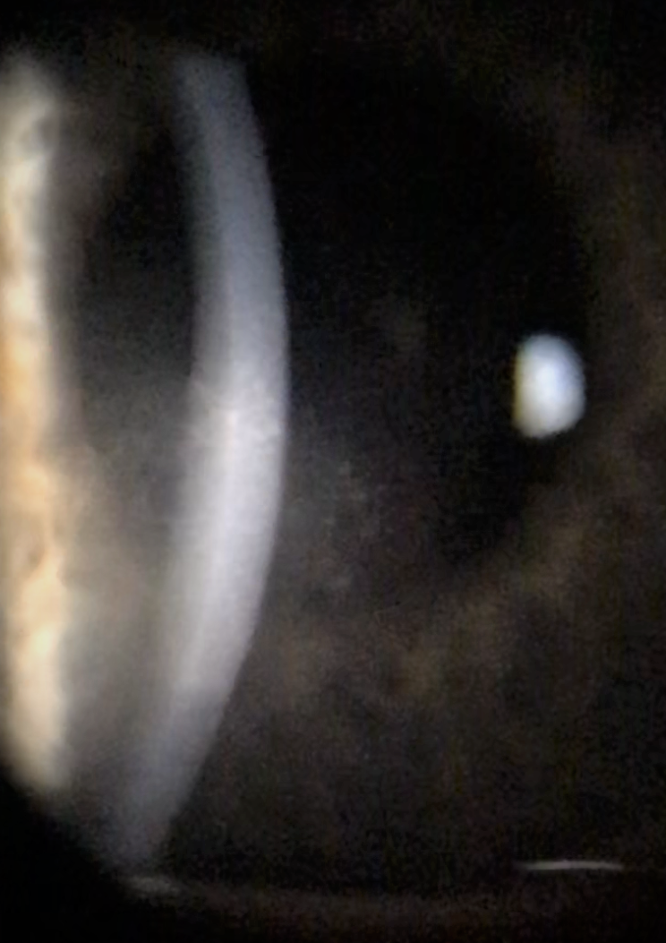
A college-aged patient came in just before final exams complaining of blurred vision in the right eye.  The eye was a little red and the vision was down to 20/40.  The cornea was swollen with subepithelial and endothelial infiltrates as shown below.  The patient has HSV disciform keratitis which requires topical steroids and oral anti-virals.  Recalcitrant or recurrent disease sometimes requires endothelial surgery to rehabilitate the cornea.  


Saturday, April 20, 2019

EPIBLEPHARON


Epiblepharon occurs when the lashes turn in  even though the eyelid position is normal. It's typically seen in children and young adults of Asian descent. Affected patients have a congenital horizontal fold of skin near the lower eyelid that pushes the eyelashes toward the eyeball.  In this case, you can see the lashes scratching the surface of the cornea.